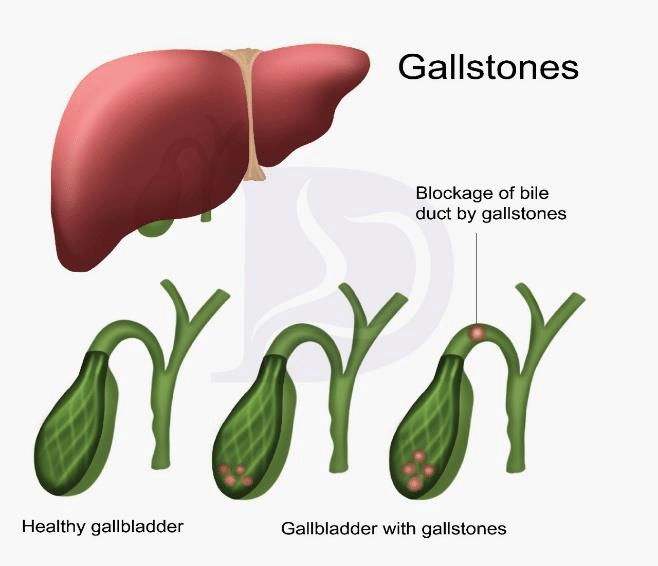
Gallstones consist of hardened fluid or mass that deposits within the gallbladder and can potentially block the bile ducts. Gallstones can range in size from a grain of sand to a golf ball!
The gallbladder is a pear-shaped organ involved in bile production that helps digest fats. It resides under the liver and is directly connected with it through a bile duct. Bile is produced in the liver and stored in the gallbladder and then passed on to the intestine for digestion of fats and oils1.
How are Gallstones formed?
Bile is composed of many chemical ingredients like salts, pigments, cholesterol, and enzymes. The gallbladder absorbs liquid from the bile, which thickens it. Crystals of cholesterol and pigments may accumulate, making the bile concentrated. These crystals continue to grow, and stone formation starts. Causes of gallstone can be:
• Increased bile secretions due to liver cirrhosis, blood disorders, and infections.
• Higher cholesterol levels
Types of Gallstones
80% of gallstones are composed of cholesterol, while the rest consists of pigments, salts, and other chemicals2.
Cholesterol gallstones: these are the most common gallstones. They are a greenish-yellow color.
Pigment gallstones: these are less-common gallstones. These are made of bilirubin so they exhibit a
darker color.
Signs and symptoms
Initially, no symptoms appear. As the condition worsens, the following may occur3:
• Pain at the right upper side of the abdomen in the liver region
• Pain in the back and shoulders
• Indigestion and upset stomach
• Heartburn, gas, vomiting, and dizziness
• Abdominal pain
• Dark urine and light-colored feces
• Yellow eyes and skin
• The gallbladder may show inflammation, and its size may double
See your doctor when
• Experiencing severe abdominal pain (colic pain)
• Yellowish skin and eyes
• High fever with chills
Diagnosis
Obtaining a detailed history is the first step in the diagnostic procress. The other steps are:
Ultrasound: A quick and straightforward method to diagnose gallstones. This study uses sound waves to display images that will detail the presence of stones.
Blood tests: Signs of infection, blockage, and inflammation can be checked through blood tests.
CT Scan: Radiographic images can be taken to visualize the presence of gallstones.
Cholescintigraphy (HIDA Scan): In this scan, the squeezing function of the gallbladder is checked with help from radiographic tracers.
Endoscopic retrograde cholangiopancreatography (ERCP): In this technique, an endoscope attached to a camera is inserted through the mouth to the small bowel, and a dye can thenbe injected in the common bile duct in order visualize the ducts and gallbladder.
Treatment of Gallstones
Smaller gallstones may clear through bile and resolve on their own.
Oral dissolution therapy: Certain medicines are used orally to break up and dissolve gallstones.
These are comprised of bile acids that help break down gallstones.
Lithotripsy: shock waves can help break gallstones into pieces through a lithotripsy machine.
Percutaneous drainage of the gallbladder: a sterile needle is inserted directly into the
gallbladder to drain the concentrated bile via a drainage tube.
Cholecystectomy
This is a surgical procedure that involves removal of the gallbladder.
What preventive measures can be taken to avoid gallstones?
• Achieve a healthy body weight, and avoid intense weight loss.
• Consume a diet high in fiber, vitamin C, mono and polyunsaturated fatty acids, vegetable
proteins, and caffeinated coffee.
• Walking, exercise, aerobic activities, and a general healthy lifestyle should be maintained.
• Limit fried foods and opt for fresh fruits and vegetables6.
Summary
Gallstones are a common disorder worldwide. Gallstones can cause severe pain and can also cause an
infection of the gallbladder or connecting ducts. Therefore, it is essential to diagnose, treat, and even go
for surgery in a timely manner to avoid serious complications. The choice of treatment can be selected
by consulting with your provider.
References:
1. Almajid AN, Sugumar K. Physiology, Bile. StatPearls. Published online September 16, 2021.
Accessed May 6, 2022. https://www.ncbi.nlm.nih.gov/books/NBK542254/
2. Tanaja J, Lopez RA, Meer JM. Cholelithiasis. Indian J Pract Pediatr. 2021;20(2):101-106.
doi:10.5005/jp/books/12296_29
3. Jones MW, Weir CB, Ghassemzadeh S. Gallstones (Cholelithiasis). StatPearls. Published online
October 9, 2021. Accessed May 6, 2022. https://www.ncbi.nlm.nih.gov/books/NBK459370/
4. McNicoll CF, Pastorino A, Farooq U, Hill CRS. Choledocholithiasis. Controv Laparosc Surg.
Published online August 31, 2021:261-267. doi:10.1007/3-540-30964-0_42
5. Hassler KR, Collins JT, Philip K, Jones MW. Laparoscopic Cholecystectomy. StatPearls. Published
online January 26, 2022. Accessed May 6, 2022.
https://www.ncbi.nlm.nih.gov/books/NBK448145/
6. Acalovschi M. Cholesterol gallstones: from epidemiology to prevention. Postgrad Med J.
2001;77(906):221. doi:10.1136/PMJ.77.906.221